Research-use-only context. This article explains the published biology of joint tissues and summarizes third-party scientific literature — most of it conducted in cultured cells or animal models. It is not medical advice, not a diagnosis, and not a treatment, therapeutic, or product claim. American Peptides products are sold strictly for in vitro laboratory research and are not for human or veterinary use.
Joint tissue degeneration is among the most extensively studied subjects in musculoskeletal research, and knee cartilage in particular is one of the field’s central model systems. This article does not offer a treatment or a protocol. Instead, it summarizes the biology as the literature describes it: what happens inside a joint as its tissues wear, why cartilage and tendons degrade, why these tissues are so slow to repair, and what directions regenerative science is exploring in the laboratory. Understanding the mechanism is the first step to reading the research intelligently.
The knee is a living tissue system
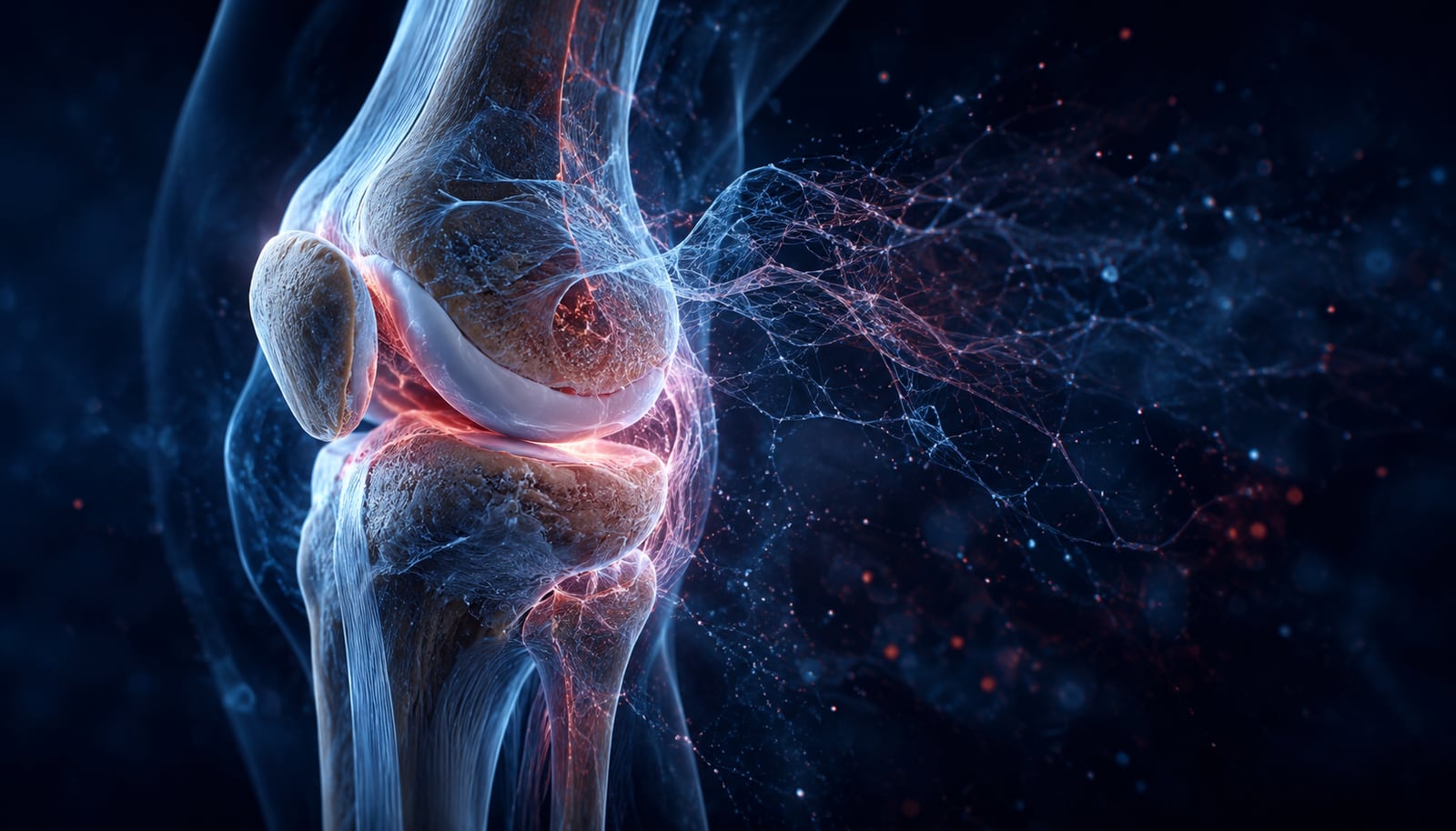
A joint is not a hinge of inert parts. The knee is a coordinated system of living tissues: a smooth layer of articular cartilage capping the ends of the bones, a lubricating synovial membrane, shock-absorbing menisci, and the tendons and ligaments that transmit and stabilize load. Every one of these is built, maintained, and remodeled by cells — and every one can fall out of balance. When researchers study “joint health,” they are really studying the biology of these tissues and the signaling that keeps them in repair.
Why cartilage breaks down
Articular cartilage is a dense matrix of type II collagen and proteoglycans (chiefly aggrecan), maintained by a sparse population of cells called chondrocytes. In a healthy joint, chondrocytes constantly balance two opposing jobs: building new matrix and clearing away old matrix. Osteoarthritis — the most-studied form of joint wear — is understood in the research literature as a breakdown of that balance. Reviews of osteoarthritis pathogenesis describe a shift toward degradation, driven by matrix-degrading enzymes such as matrix metalloproteinases (MMPs) and aggrecanases and amplified by inflammatory signaling molecules [1,2]. The outcome is a gradual loss of the collagen-and-proteoglycan scaffold that gives cartilage its spring and resilience.
Those inflammatory signals are real and measurable, and reading them is its own discipline — see the explainer on inflammation biomarkers.
Why cartilage repairs so slowly
Here is the biological catch that makes cartilage degeneration such a persistent research problem: mature articular cartilage has almost no capacity to repair itself. Unlike skin or muscle, it is avascular (no blood supply), aneural (no nerves), and populated by very few cells. With no blood-borne repair cells reaching the tissue and a low resident cell density, the intrinsic healing response is minimal — a limitation reviewed at length in the cartilage tissue-engineering literature [3]. This is exactly why so much regenerative research targets cartilage: the tissue that most needs repair is the one least able to manage it on its own.
Tendons: the other half of the story
Cartilage is not the only tissue studied in joint degeneration. Tendons — the collagen-rich cords that connect muscle to bone — are a second major research focus, and their biology follows a similar theme. Tendons are strong but relatively slow to heal; tendon healing proceeds through overlapping inflammation, repair, and remodeling phases, and the repaired tissue often never fully regains the highly organized collagen architecture of the original [4,5]. When healing repeatedly fails to keep pace with load, the result is tendinopathy — described in the research literature not as simple “inflammation” but as a failed or disordered healing response of the tendon matrix [6]. Once again the through-line is the same: the tissue’s own repair machinery is the bottleneck.
The role of signaling
Whether the tissue is cartilage or tendon, repair is orchestrated by signaling molecules — the chemical messages that tell cells when to divide, migrate, build matrix, or stand down. Many of those messengers are themselves peptides: short chains of amino acids that bind receptors and switch cellular programs on and off. (For the fundamentals, see what research peptides are and how signaling peptides work.) That is why peptides have become such an active area of tissue-repair research — they sit at the exact control points the biology above depends on.
What regenerative research is exploring
A number of signaling peptides have been studied in the context of tissue repair. It is important to frame this honestly: the large majority of that work is preclinical — conducted in cultured cells or animal models — and does not establish outcomes in people. With that caveat, three of the most-studied compounds in the regenerative literature are:
- BPC-157 — a peptide studied in animal models of tendon, ligament, and gut-tissue injury. The published evidence, and its limits, is reviewed in BPC-157 research: what the studies show.
- TB-500 / Thymosin β4 — corresponds to the actin-binding region of a protein studied for decades in cell migration and tissue repair. See TB-500 research: what the studies show.
- GHK-Cu — a copper-binding tripeptide with a deep literature in matrix remodeling and gene expression. See GHK-Cu research: what the studies show.
Across all three the pattern is consistent: mechanistically interesting, biologically coherent, and still largely preclinical. None is an approved drug for joint or tendon repair. The value of this literature is descriptive — it maps where the science genuinely stands.
Recovery itself is increasingly treated as a measurable variable in research; that shift is covered in the science of recovery, why recovery is becoming a performance metric, and what the research says about sleep and cellular repair.
Reading the research responsibly
Two principles keep this kind of reading honest. First, preclinical is not proof. A promising result in a rodent tendon model is a reason for more research, not a conclusion about people. Second, the label matters. The compounds discussed here are sold strictly for laboratory research — see what “research use only” actually means — and evaluating any research compound starts with verifying its identity and purity through a Certificate of Analysis. Researchers surveying the tissue-repair literature can browse the compounds referenced above in the tissue-research catalog, and the full plain-language library lives in the research education hub.
Frequently Asked Questions
How does the research literature explain age-related joint tissue wear?
Research on osteoarthritis describes a gradual shift in cartilage from matrix-building toward matrix-degradation, driven by enzymes and inflammatory signaling. Because mature cartilage is avascular and repairs poorly, that wear accumulates over time. This is a description of published biology, not medical advice.
Does the literature describe cartilage as capable of self-repair?
Mature articular cartilage has very limited intrinsic repair capacity because it is avascular, aneural, and low in cells. That limitation is precisely why cartilage is a major focus of regenerative research.
How does the literature distinguish cartilage degeneration from tendon pathology?
Cartilage is the smooth matrix capping the bone ends inside the joint; tendons are the collagen cords connecting muscle to bone. Tendinopathy is described in the literature as a disordered healing response of the tendon matrix, distinct from cartilage wear.
What does the research literature say about peptides studied in joint and tendon tissue models?
Several signaling peptides — including BPC-157, TB-500, and GHK-Cu — have been studied in preclinical tissue-repair models. The evidence is largely animal- and cell-based, and none is an approved therapy. See the linked study reviews for what each literature actually reports.
Citations
- “Osteoarthritis pathogenesis: a review of molecular mechanisms.” Calcif Tissue Int. 2014. PubMed: PMID 25311420
- “Recent Updates of Diagnosis, Pathophysiology, and Treatment on Osteoarthritis of the Knee.” Int J Mol Sci. 2021. PubMed: PMID 33807695
- “Recent advances in hydrogels for cartilage tissue engineering.” Eur Cell Mater. 2017 (reviews cartilage’s limited intrinsic repair capacity). PubMed: PMID 28138955
- “Biology and physiology of tendon healing.” Joint Bone Spine. 2024. PubMed: PMID 38307405
- “Tendon basic science: Development, repair, regeneration, and healing.” J Orthop Res. 2015. PubMed: PMID 25764524
- “Tendinopathy.” Nat Rev Dis Primers. 2021. PubMed: PMID 33414454
This article is for laboratory research reference only. American Peptides products are sold strictly for in vitro research. Not for human consumption.
Related research
- The Biology of Sleep and Cellular Repair: What the Research Literature Describes
- The Future of Healthspan Research: A Research Overview
- Amino Acid Signaling and Receptor Biology: A Research Primer
- What Is Selank? A Research Overview of the Peptide and Its Studied Pathways
- What Is Semax? A Research Overview of the Peptide and Its Studied Pathways
Compliance Notice: American Peptides products are sold strictly for laboratory and academic research purposes only. They are not intended for human or veterinary consumption, diagnosis, treatment, or prevention of any disease. All content on this page is educational in nature and does not constitute medical advice or product claims. Researchers are responsible for handling these compounds in accordance with their institution’s safety protocols and applicable laws.